sexta-feira, junho 10, 2011

Campo magnético "afina" o sangue para prevenir ataques cardíacos

09/06/2011


Redação do Diário da Saúde


Campo magnético


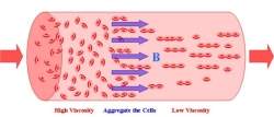
     O campo magnético polariza as células vermelhas do sangue, fazendo com que elas se liguem a cadeias curtas, melhorando a circulação do sangue.[Imagem: Temple University]

Afinar o sangue

Se o sangue de uma pessoa se torna muito espesso, ele pode danificar os vasos sanguíneos e aumentar o risco de ataques cardíacos.

Mas um físico da Universidade Temple (EUA) descobriu que pode-se diluir o sangue humano submetendo-o a um campo magnético.

O físico Rongjia Tao foi pioneiro no uso de campos elétricos e magnéticos para diminuir a viscosidade do óleo em motores e dutos.

Agora, ele está usando os mesmos campos magnéticos para afinar o sangue humano no sistema circulatório.

Magnetismo no sangue

Como os glóbulos vermelhos contêm ferro, Tao foi capaz de reduzir a viscosidade do sangue de uma pessoa entre 20 e 30 por cento, sujeitando-o a um campo magnético de 1,3 Tesla por cerca de um minuto.

Esse campo magnético tem aproximadamente a mesma intensidade da usada em um exame de ressonância magnética (MRI).

Depois de testarem diversas amostras de sangue em laboratório, os pesquisadores descobriram que o campo magnético polariza as células vermelhas do sangue, fazendo com que elas se liguem a cadeias curtas, melhorando a circulação.

Como essas cadeias são maiores do que as células do sangue individuais, elas fluem para o centro, reduzindo o atrito contra as paredes dos vasos sanguíneos.

Os efeitos combinados reduzem a viscosidade do sangue, ajudando-o a fluir mais livremente.

Viscosidade do sangue

Quando o campo magnético é retirado, a viscosidade inicial do sangue lentamente retorna, mas em um período de várias horas.

A técnica é conhecida como magneto-reológica e, sendo reversível, aponta para uma possibilidade de uso em situações críticas ou na prevenção de ataques cardíacos - os campos magnéticos podem ser reaplicados e a viscosidade reduzida novamente.

"Selecionando uma intensidade do campo magnético e uma duração de pulso adequadas, podemos controlar o tamanho das cadeias agregadas de hemácias, controlando, portanto, a viscosidade do sangue," disse Tao.

Método do campo magnético

Atualmente, o único método para diluir o sangue é através de drogas, como a aspirina. No entanto, essas drogas frequentemente produzem efeitos colaterais indesejados.

Tao afirma que o método do campo magnético não é apenas mais seguro, mas também pode ser aplicado repetidas vezes.

Ele também acrescentou que a redução da viscosidade não afeta a função normal das células vermelhas do sangue.

O pesquisador alerta que mais estudos serão necessários e que ele espera conseguir transformar esta tecnologia em uma terapia para prevenir doenças cardíacas, que seja aceita pelas autoridades de saúde.

Fonte: http://www.diariodasaude.com.br/news.php?article=campo-magnetico-afina-sangue&id=6588&nl=nlds.

terça-feira, abril 26, 2011

Medical Imaging International — MII April 2011

Two Acceleration Methods Allow For Much Faster MRI Brain Scans

An international team of physicists and neuroscientists has reported a breakthrough in magnetic resonance imaging (MRI) that allows brain scans more than seven times faster than currently possible. In an article published December 20, 2011, in the journal PloS ONE, a University of California (UC) Berkeley (USA; http://berkeley.Edu), physicist and colleagues from the University of Minnesota (Twin Cities [Minneapolis-St. Paul], USA; www.umn.edu) and Oxford University (UK; www.ox.ac.uk) described two developments that allow full three-dimensional (3D) brain scans in less than half a second, instead of the typical two to three seconds.
“When we made the first images, it was unbelievable how fast we were going,” said first author Dr. David Feinberg, a physicist and adjunct professor in UC Berkeley’s Helen Wills Neuroscience Institute and president of the company Advanced MRI Technologies (Sebastopol, CA, USA). “It was like stepping out of a prop plane into a jet plane. It was that magnitude of difference.”
For neuroscience, in particular, fast scans are vital for capturing the dynamic activity in the brain. “When a functional MRI [fMRI] study of the brain is performed, about 30 to 60 images covering the entire 3D brain are repeated hundreds of times like the frames of a movie but, with fMRI, a 3D movie,” Dr. Feinberg said. “By multiplexing the image acquisition for higher speed, a higher frame rate is achieved for more information in a shorter period of time.”
“The brain is a moving target, so the more refined you can sample this activity, the better understanding we will have of the real dynamics of what’s going on here,” added Dr. Marc Raichle, a professor of radiology, neurology, neurobiology, biomedical engineering, and psychology at Washington University in St. Louis (MO, USA; http://wustl.edu), who has followed Dr. Feinberg’s work.
Because the technique works on all modern MRI scanners, the impact of the ultrafast imaging technique will be immediate and widespread at research institutions worldwide, according to Dr. Feinberg. In addition to greatly advancing the field of neural imaging, the discovery will have an immediate impact on the Human Connectome Project (http://humanconnectomeproject.org), funded in 2010 by the US National Institutes of Health (NIH; Bethesda, MD, USA) to map the connections of the human brain through fMRI and structural MRI scans of 1,200 healthy adults.
“At the time we submitted our grant proposal for the Human Connectome Project, we had aspirations of acquiring better quality data from our study participants, so this discovery is a tremendous step in helping us accomplish the goals of the project,” said Dr. David Van Essen, a neurobiologist at Washington University and coleader of the project. “It’s vital that we get the highest quality imaging data possible, so we can infer accurately the brain’s circuitry – how connections are established, and how they perform.”
The faster scans are made possible by combining two technical improvements devised in the past decade that separately increased scanning speeds two to four times over what was already the fastest MRI technique, echo planar imaging (EPI). Physical limitations of each method prevented further speed improvements, “but together their image accelerations are multiplied,” Dr. Feinberg said. The researchers can now obtain brain scans considerably faster than the time reductions reported in their study and many times faster than the capabilities of current machines.
Nearly 20 years ago, however, a new type of MRI called functional MRI (fMRI) was developed to highlight areas of the brain using oxygen, and thus seemingly engaged in neuronal activity, such as thinking. Utilizing EPI, fMRI vividly differentiates oxygenated blood funneling into working areas of the brain from deoxygenated blood in less active areas.
With EPI, a single pulse of radio waves is used to excite the hydrogen atoms, but the magnetic fields are rapidly reversed several times to elicit about 50 to 100 echoes before the atoms settle down. The multiple echoes provide a high-resolution image of the brain. In 2002, Dr. Feinberg proposed using a sequence of two radio pulses to obtain twice the number of images in the same amount of time. Called simultaneous image refocusing (SIR) EPI, it has proved useful in fMRI and for 3D imaging of neuronal axonal fiber tracks, though the improvement in scanning speed is limited because with a train of more than four times as many echoes, the signal decays and the image resolution decreases.
Another acceleration advance, multiband excitation of several slices using multiple coil detection, was proposed in the United Kingdom at about the same time by Dr. David Larkmann for spinal imaging. The technique was used for fMRI by Dr. Steen Moeller and colleagues at the University of Minnesota. This technique, too, had limitations, mainly because the multiple coils are relatively widely spaced and cannot differentiate very closely spaced images.
The ability to scan the brain in under 400 ms moves fMRI closer to electroencephalography (EEG) for capturing very rapid sequences of events in the brain. The development will affect general fMRI as well as diffusion imaging of axonal fibers in the brain, both of which are needed to achieve the main goal of the Human Connectome Project. Diffusion imaging reveals the axonal fiber networks that are the main nerve connections between areas of the brain, while fMRI shows which areas of the brain are functionally connected, meaning, which areas are active together or sequentially during various activities.


Fonte: http://www.mydigitalpublication.com/.

novo-2

sábado, abril 23, 2011

SUS ganhará aparelho capaz de destruir câncer


O aparelho é uma tecnologia inovadora, resultante da fusão do ultrassom de alta intensidade com a ressonância magnética

O SUS irá ganhar um novo equipamento com capacidade para destruir tumores cancerígenos. O aparelho chamado de High Intense Focus Ultrassound (Hifu) será disponibilizado pelo Instituto do Câncer do Estado de São Paulo (Icesp). E, é uma tecnologia inovadora, resultante da fusão do ultrassom de alta intensidade com a ressonância magnética.

Pioneiro na América do Sul, o novo procedimento será utilizado, inicialmente, para tratar miomas e metástases ósseas, mas a ideia é ampliar seu uso para outras áreas da oncologia. O investimento para aquisição do equipamento foi de R$ 1,5 milhão.

O aparelho permitirá investigar novas terapias que, aliando o ultrassom à ressonância magnética, viabilizarão o tratamento de tumores sem a necessidade da realização de cortes e cirurgia ou de internação. Por não ser invasivo, o método, que dura aproximadamente duas horas, permite que o paciente realize o procedimento consciente, permanecendo acordado e podendo voltar para casa no mesmo dia.

O Hifu concentra até 1.000 feixes de energia ultrassônica com extrema precisão em um tumor no interior do corpo. Cada feixe passa através do corpo sem causar lesão, mas, quando convergem para o ponto selecionado, elevam a temperatura nesse local. A ressonância magnética serve para localizar e direcionar essa energia precisamente no tumor, de forma interativa e em tempo real, fornecendo imediata confirmação da eficácia da terapia.

Além disso, estão sendo desenvolvidos no Icesp tratamentos que possibilitam a liberação de drogas quimioterápicas, em que nanopartículas com elevadas concentrações de medicamentos (o que pode ser altamente tóxico ao organismo, inviabilizando sua aplicação intravenosa), são injetadas e liberadas apenas no tumor, a partir do calor produzido pelo aparelho.

Os pacientes que se beneficiarão da novidade integrarão os protocolos de pesquisa clínica do Icesp. Além de esta ser uma novidade na área oncológica, a aquisição do equipamento estabelece inúmeras possibilidades e caminhos no ambiente de pesquisa.

Isto representa um grande avanço não apenas para os pacientes do SUS, como também para a instituição, que se reafirma como referência na área de investigação e tratamento do câncer. Ganha, também, o país, que passa a ser reconhecido por sua produção científica e desenvolvimento de novos protocolos e tratamentos.


Fonte: http://www.saudebusinessweb.com.br

Página Inicial

sexta-feira, abril 22, 2011

Biópsia Virtual usando espectroscopia de MR pode permitir um diagnóstico mais precoce de transtorno cerebral em atletas.


Em um estudo com ex-atletas profissionais, pesquisadores descobriram que uma técnica de imagem especializada chamada espectroscopia por ressonância magnética (RM) podem ajudar a diagnosticar traumática crônica en espongiforme bovina (CTE), uma doença causada por traumatismo craniano repetitivo, que atualmente só pode ser diagnosticada na autópsia .
Os resultados do estudo foram apresentados em dezembro de 2010 na reunião anual da Radiological Society of North America (RSNA), realizada em Chicago, IL, EUA. "Os efeitos devastadores das lesões cerebrais sofridas pelos jogadores de futebol profissional que repetidamente sofreram abalos e traumatismos cranianos subconcussive durante suas carreiras colocou os holofotes sobre CTE", disse Alexander P. Lin, PhD, principal investigador do Centro de Clínica Espectroscopia na Brigham and Women's Hospital (Boston, MA, EUA; www.mgh.harvard.edu).
Segundo os Centros de Controle de Doenças dos EUA (CDC, Atlanta, GA, EUA), estima-se 3,8 milhão de esportes e recreação abalos relacionados ocorrem nos Estados Unidos a cada ano. Além disso, os abalos subclínica - lesões que não podem ser diagnosticados como concussões, mas têm efeitos semelhantes - vão frequentemente unrecognized. Estudos têm demonstrado que indivíduos que sofrem trauma crânio-encefálico repetitivos são mais propensos a ter problemas em curso, de dano cerebral permanente de incapacidade a longo prazo.
CTE é uma doença degenerativa do cérebro causada por traumas cerebrais repetidas e marcada por um acúmulo de proteínas anormais no cérebro. CTE tem sido associada à dificuldade de memória, comportamento impulsivo e errático, depressão e, finalmente, a demência. "Traumatismo craniano cumulativa invoca mudanças no cérebro, que ao longo do tempo pode resultar em um declínio progressivo da memória e funções executivas em alguns indivíduos," diz Lin. "MRS pode nos fornecer, detecção não invasiva precoce do CTE antes de mais danos ocorre, permitindo assim uma intervenção precoce".
No estudo de Lin, realizado em colaboração com a Universidade de Boston Center for the Study of Traumatic Encefalopatia (CSTE; www.bu.edu/ CSTE), cinco atletas do sexo masculino profissional aposentado do futebol, wrestling e boxe com o CTE suspeitos e cinco de idade e tamanho controles pareados entre as idades de 32 e 55 foram examinados com a MRS. Na MRS, por vezes referido como "biópsia virtual", um poderoso campo magnético e ondas de rádio são usadas para extrair informações sobre compostos químicos no corpo, usando um scanner de ressonância magnética clínica.
Os resultados revelaram que, em comparação com os cérebros de pacientes do grupo controle, o cérebro dos ex-atletas com CTE suspeitos tinham aumentado os níveis de colina, um nutriente membrana celular que sinaliza a presença de tecido danificado e glutamato / glutamina, ou GLX. MRS também revelou alterações nos níveis de ácido gama-aminobutírico (GABA), aspartato e glutamato no cérebro de ex-atletas. "Ao nos ajudar a identificar os neuroquímicos que podem desempenhar um papel no CTE, este estudo tem contribuído para o nosso entendimento da fisiopatologia da doença," diz Lin.
Por exemplo, os aminoácidos e neurotransmissor glutamato está envolvido na maioria dos aspectos da função cerebral normal e deve estar presente nos lugares certos e na concentração certa para que o cérebro para ser saudável - muito ou pouco pode ser prejudicial. Resultados de estudos neuropathologic CSTE de jogadores de futebol aposentado e outros atletas levaram a mudanças consideráveis ​​na NFL ([dos EUA] da National Football League), bem como esportes universitários e jovens.

Fonte: http://www.mydigitalpublication.com/publication/?i=62726

medicalimaging.com

sexta-feira, março 04, 2011

Detecção de infarto do miocárdio em pacientes usuários de cocaína .

Detecção de infarto do miocárdio através de ressonância magnética cardiovascular e angiotomografia coronária em pacientes usuários de cocaína com história de dor torácica após seu uso

INTRODUÇÃO:
A cocaína é a terceira droga ilícita mais comumente utilizada nos Estados Unidos e a principal responsável pelo atendimento de pacientes usuários de drogas em serviços de emergência médica.
A queixa mais comum na entrada da emergência é a dor torácica, referida em 40% dos casos. Além disso, o uso crônico leva a piora da hipertensão, hipertrofia ventricular esquerda e acelera a aterosclerose. A ressonância magnética cardiovascular é um excelente método para avaliação da morfologia e função ventricular, com excelente reprodutibilidade, e atualmente considerada padrão ouro.
A angiotomografia coronária é um método diagnóstico em ascensão, permitindo a detecção de DAC obstrutiva e não obstrutiva, acrescentando informação para a estratificação de risco cardiovascular. O objetivo desse estudo foi avaliar a eventual presença de infarto prévio em pacientes jovens (18 a 40 anos) usuários de cocaína, que apresentavam dor torácica, através da detecção de fibrose miocárdica por exame de ressonância magnética cardiovascular. O objetivo secundário foi avaliar alterações parietais e obstruções das coronárias desses pacientes por angiotomografia coronária.
MÉTODOS:
Avaliamos 24 pacientes usuários de cocaína (nas formas inalatória, injetável ou crack) que apresentavam dor torácica freqüente e de longa duração relacionada ao seu uso. Esses pacientes realizaram a angiotomografia coronária e a ressonância magnética cardiovascular.
A angiotomografia coronária avaliou o escore de cálcio e árvore coronária por segmentos, e a ressonância magnética cardiovascular avaliou dimensões, volumes e função ventricular, bem como a eventual presença de realce tardio miocárdico.
RESULTADOS: Foram estudados 24 pacientes, 22 homens, com idade média de 29,7 anos (18 a 40 anos). A grande maioria dos pacientes (79%) fazia uso de cocaína inalatória de forma freqüente e 71% dos pacientes já haviam usado crack. O escore de cálcio foi positivo em apenas um paciente [54 (Agatston) e 56 (volume)].
Nenhum dos pacientes apresentou redução luminal significativa. Dos segmentos coronários avaliados, apenas um paciente apresentou placas calcificadas na artéria descendente anterior, nos segmentos proximal e médio. A análise da função ventricular global através da fração de ejeção (FE), volume diastólico final (VDF), volume sistólico final (VSF), e massa ventricular foi considerada normal em 100% da amostra. As médias da FE, VDF e VSF foram 60,7%, 147,7 ml e 59,1 ml, respectivamente.
O índice cardíaco foi considerado normal em todos os pacientes, com média de 2,9. Nenhum paciente apresentou hipertrofia miocárdica. A análise da função ventricular segmentar através da análise dos 17 segmentos foi normal em todos os pacientes. Nenhum paciente apresentou realce tardio indicativo de fibrose miocárdica.
CONCLUSÃO:
A ressonância magnética cardiovascular não demonstrou, na população estudada, a presença de realce tardio miocárdico indicativo de fibrose, em indivíduos jovens e de baixo risco para doença arterial coronária, e com episódios de dor precordial prolongada durante ou após o uso de cocaína.
A tomografia computadorizada corroborou o perfil de baixo risco clínico, demonstrando a ausência de ateromatose coronária na grande maioria (96%) dos indivíduos estudados. Apenas uma pequena porcentagem (4%) dos indivíduos apresentou ateromatose coronária discreta, sem obstruções significativas
Assessment of myocardial infarction by cardiovascular magnetic resonance and computed tomography angiography in patients with cocaine-associated chest pain
Abstract in English
INTRODUCTION: Cocaine is the third most commonly used illicit drug in the United States and the leading cause of emergency department visits among drug users.
Chest pain is the most common cocaine-related presentation, being reported in 40% of patients. Its chronic use causes hypertensive crises, myocardium hypertrophy and accelerates the process of atherosclerosis.
Cardiovascular magnetic resonance provides an accurate assessment of cardiac morphology and ventricular function with excellent reproductibility, and it is considered the gold standard method.
Computed tomography angiography has emerged as a powerful tool to evaluate patients with suspected coronary artery disease at the same time that it helps in the prognostic assessment of the patient.
The purpose of this study was to evaluate the incidence of previous myocardial infarction among young cocaine users (18 to 40 years) with chest pain related with the use of the drug by the assessment of myocardial fibrosis through cardiovascular magnetic resonance. Secondarily, was also meant the evaluation of the coronary tree by the computed tomography angiography.
METHODS:
We studied 24 cocaine users (crystalline, powder or granular forms) that frequently complained about chest pain related to the use of the cocaine.
These patients underwent computed tomography angiography with assessment of calcium score and the evaluation of the segmented coronary arteries, and cardiovascular magnetic resonance to assess dimensions, volumes and ventricular function of the heart, and the presence of myocardial fibrosis.
RESULTS:
We studied 24 patients (22 male), mean age of 29.7 years. Most of the patients (79%) had frequently used inhalatory cocaine, 71% of them had also used the crack cocaine form.
The calcium score turned out to be positive in only one patient [54 (Agatston) and 56 (volume)]. None of them showed significant coronary stenosis.
Among the coronary segments evaluated, only one patient had calcified plaques at the anterior descending coronary artery (proximal and medium segments).
The global analysis of the left ventricular function assessed by the ejection fraction (EF), end diastolic volume (EDV), end sistolic volume (ESV) and ventricular mass were considered normal in 100% of the patients. Mean EF, EDV and ESV were 60.7%, 147.7mL and 59.1mL respectivelly.
Cardiac index was normal in all patients. None of the patients showed myocardial hypertrophy. Assessment of regional ventricular function by the evaluation of 17 segments was normal in all patients. None of the patients showed myocardial delayed-enhancement, indicative of myocardial fibrosis.
CONCLUSION:
Cardiovascular magnetic resonance did not detect the presence of delayed enhancement indicative of myocardial fibrosis among young cocaine users with low cardiovascular risk with complaints of chest pain during or after cocaine abuse. Computed tomography angiography confirmed low cardiovascular risk of these patients, since most of them (96%) had no atherosclerosis detected by this exam. Only one patient (4%) had coronary atherosclerosis detected, without significant coronary stenosis
www.teses.usp.br

Fonte:http://www.radiology.com.br/materias/rad_materias.asp?flag=1&id_materia=945.

sábado, janeiro 29, 2011

FDA americano avalia riscos de contraste para RM da Bayer

Os especialistas americanos foram alertados para ficarem atentos aos efeitos potenciais como sérios danos à pele causados pelo contraste para ressonância da Bayer AG. A Food and Drug Administration em documento divulgado esta semana, indicam este contraste para uma lista de outras medicações semelhantes, potenciais causadoras de fibrose sistêmica nefrogênica (NSF), uma doença potencialmente fatal de pele.
O produto da Bayer, o gadobutrol, é uma versão fortificada de uma classe de agentes à base de gadolineo, ou GBCAs, utilizado em exames de ressonância magnética como meio de contraste.
“Devido ao risco de Fibrose Sistêmica Nefrogênica associado à doses elevadas dos GBCAs, uma potencial overdose do gadobutrol inplica em riscos desta patologia para os pacientes” indica o relatório prévio a respeito da questão divulgado pelo FDA.
A empresa têm solicitado ao FDA para aprovação da comercialização de gadobutrol utilizado em Ressonancias do Sistema Nervoso Central. Cinco outras drogas foram aprovadas para uso com efeito intermediário ao gadobutrol.
Em setembro do ano passado, o FDA havia divulgado um alerta específico sobre os GBCAs a cerca do risco de nefrose sistêmica em pacientes portadores de patologias renais. Contrastes como o Magnevist da Bayer, Covidiens Optimark e Omniscan,produzido pela GE Healthcare, foram identificados comop de alto risco em relação à outros GBCAs aprovados.
A própria Bayer relatou 10 casos de Nefrose Sistêmica desde que o Gadobutrol foi aprovado na Europa em 1998, comentou o staff do FDA. Dois pacientes receberam somente gadobutrol enquanto os outros fizeram uso de outros contrastes GBCAs.Cerca de 5 milhões de pacientes foram expostos ao gadobutrol sob o nome de Gadovist.
Os dados preliminares sugeriam que os riscos do gadobutrol para NSF eram similares ou menores do que os outros agentes, comenta o staff da FDA. Os revisores da agência também disseram sobre a efetividade da droga.
A Bayer se compromete em boletim divulgado junto à FDA para alertar no próprio medicamento sobre a intensidade do contraste e os riscos potenciais, para prevenção de eventuais overdoses.
Veja um sumário desta discussão em 
http://bit.ly/gDbiy5.
Baseado em matéria divulgada por Lisa Richwine no AuntMinnie.com

http://www.auntminnie.com/index.aspx?sec=sup&sub=mri&pag=dis&ItemID=93911

domingo, janeiro 16, 2011

Intraosseus and extraosseus juxtaarticular calcification: Osteopoikilosis with synovial osteochondromatosis - an association

 
Case Report
Parangama Chatterjee1, Jyoti Sureka1, Elizabeth Joseph1, Sniya Sudhakar1, Samuel Chittaranjan2
Radiology Case. 2009 Mar; 3(3):1-5 :: DOI: 10.3941/jrcr.v3i3.138

1. Department of Radiodiagnosis and Imaging, Christian Medical College, Vellore, Tamil Nadu, India
2. Department of Orthopaedics, Christian Medical College, Vellore, Tamil Nadu, India

Osteopoikilosis presents as round or ovoid sclerotic lesions with an appearance like enostosis on pathology. Synovial osteochondromatosis occurs due to cartilaginous metaplasia with synovial villous proliferation with calcified nodules in proximity to joints. A case of osteopoikilosis associated with synovial osteochondromatosis is described. Intraosseus and juxta osseus sclerotic bone lesions were identified on radiographs and computed tomography in a patient with knee pain. The association of osteopoikilosis with synovial osteochondromatosis is rare and to our knowledge has received little attention in the literature.

INTRODUCTION
Osteopoikilosis is a hereditary condition. The hallmark of this condition is numerous discrete or confluent round or ovoid calcific or ossific densities often with minimally spiculated margins, in bones, often in proximity to joints (
1). These are often incidentally discovered and pathologically represent bone islands. They are occasionally associated with cutaneous lesions as well as other osteosclerotic disorders (2).
Synovial osteochondromatosis is a rare benign monoarticular arthropathy. The inciting stimulus which results in the rapid development of this synovial metaplastic process is unknown (
3). An embryonic rest has been hypothesised to be the causative factor for the disease (4, 5). Trauma has also been identified as a precipitating factor (4, 6). A neoplastic basis for the cartilaginous metaplasia within the synovial villi is considered by some authors (3, 4, and 5). Although malignant transformation has been reported occasionally (4, 7, and 8) it is controversial as to whether or not chondrosarcomas actually originate in this entity. Synovial chondromatosis most commonly involves the knee, elbow, and hip in young adults (6, 9). The association of osteopoikilosis with synovial osteochondromatosis is rare and to our knowledge has received little attention in the literature.
CASE REPORT
A 25 year old man presented to the orthopaedics department in our institution with right knee pain and difficulty in walking, progressively increasing for 5 years. He also gave a history of fall, which happened 1 month before the onset of pain. There was no history of tingling or numbness or significant family history. Outside MRI showed tears of both menisci on the right, longitudinal tear of the lateral meniscus and radial tear of the medial meniscus, along with a tear of the lateral collateral ligament of the right knee. The MR also showed nodular lesions in relation to the knee which were hypointense on all sequences, few of which were intra osseous and few were extra osseous. (Figures 1a-c) The patient did not have any other complaints. Examination of the right lower limb revealed wasting of the quadriceps. Rest of the clinical examination was normal. His laboratory values including erythrocyte sedimentation rate, rheumatoid factor, C-reactive protein, uric acid, serum calcium and parathyroid hormone levels were normal.
He was referred to the radiology department for a radiograph of the knee joint, which showed mild juxta-articular osteopenia and multiple small discrete, round, oval, dense calcific lesions in a symmetric distribution (
Fig. 2

). These lesions were distributed more in the epiphysis and metaphysis of the femur distally (Fig. 3  ). An AP radiograph of the pelvis did not show any abnormality. All findings were suggestive of osteopoikilosis. There were also few intra articular soft tissue density lesions with peripheral calcifications. A CT scan done thereafter confirmed the findings. (Fig. 4  a and 4c).After the diagnosis, the patient was re-evaluated, and any overlooked pathology was searched. There was no pain or swelling of the joints, no muscle contractures, no soft tissue or skin changes.
He underwent an arthroscopic proceed open biopsy of the lateral condyle of the right femur to exclude osteoblastic metastases. Arthroscopy revealed typical features of proliferative frond like synovial disease with associated loose bodies as seen in synovial chondromatosis. A synovial debridement with removal of the loose bodies was done. These were however not sent for histopathological analysis by the operating surgeon in order to reduce patient expenses and because the arthroscopic appearances were typical. Microscopic histopathology revealed few thickened trabeculae merging with normal surrounding bone with no evidence of metastases, which can be seen with osteopoikilosis. We do not have archived pathological images as the femoral condyle biopsy was done to exclude osteoblastic metastases. Images were not archived as it was a negative study for the diagnostic question i.e. metastases.

Osteopoikilosis, also called osteopathia condensans disseminata, or "spotted bone disease" is an incidentally detected disorder characterized by an abnormality in enchondral bone maturation. There is no gender predilection, men and women are equally affected (1, 9). It is usually inherited as an autosomal dominant condition. Osteopoikilosis results in multiple small densities of oval or rounded shape that are symmetrically distributed within the metaphyses and epiphyses of the long bones, in periarticular osseous regions. On histology these foci are formed by dense trabeculae of spongious bone, occasionally forming a nidus without communication with bone marrow. These appear in childhood and persist thereafter (2, 5, 10). Sites of predilection include phalanges (100%), carpal bones (97.4%), metacarpals (92.5%), foot phalanges (87.2%), metatarsals (84.4%), tarsal bones (84.6%), pelvis (74.4%), femur (74.4%), radius (66.7%), ulna (66.7%), sacrum (58.9%), humerus (28.2%), tibia (20.5%), and fibula (2.8%) in one study of 4 families. The ribs, skull and vertebrae are spared (2).
Some patients have associated connective tissue nevi called dermatofibrosis lenticularis disseminate, this is called Buschke-Ollendorff syndrome (
11). Osteopoikilosis is also associated with keloid formation, dwarfism, spinal stenosis, dystocia, melorrheostosis, tuberous sclerosis and scleroderma (6, 7, and 13). Mixed sclerosing bone dystrophy comprises osteopathia striata and melorheostosis along with osteopoikilosis (2).
Differential diagnoses include osteoblastic metastasis, tuberous sclerosis and mastocytosis (
1, 9). Blood investigations like alkaline and acid phosphatase and bone scan are typically normal (1, 2, 14).
The diagnosis of primary synovial osteochondromatosis is based on the rapid onset of monoarticular pathology in a young adult and on the gross histopathological appearance (
3). The typical features are ectopic chondroid matrix formation under the synovium, synovial hyperplasia, multiple small loose bodies and the absence of articular cartilage destruction. However secondary synovial metaplasia occurs in the more common traumatic and degenerative joint disorders (3, 5, and 6). Radiographs may or may not reveal calcific densities.
Synovial chondromatosis progresses through various stages of activity (
4, 13). In the acute stage, the entire joint synovium is hypertrophied and hyperemic with numerous foci of cartilage formation. Small cartilaginous fragments are extruded into the joint and dystrophic calcification may occur. (3,6) During the intermediate stage, the acute synovial reaction gradually subsides (7,10,13, 15)The free cartilaginous loose bodies undergo degenerative, proliferative (enlargement on multiplication), or resorptive changes, depending on stresses peculiar to the joint involved and location within the joint (4,7). Endochondral bone formation may occur but requires a blood supply and is confined to loose bodies with a pedicle or to free loose bodies that have regained a synovial attachment (4,9).
In the late stages of the disease, the generalized synovial reaction reverts to normal. Secondary osteoarthritis results from the presence of loose bodies within the joint in chronic cases (
6, 8, 9). This may cause localized secondary synovial chondromatosis, as well as osteochondritic loose bodies formed by detached osteophytes. Therefore, in older cases of long duration only a presumptive diagnosis of primary synovial chondromatosis can be made based on the history and the number and character of the loose bodies.
Synovial chondrosarcoma has to be considered in the differential diagnosis of synovial chondromatosis since both lesions may exhibit synovial calcification and atypical multinucleated cartilage cells (
9, 13, and 16). The diagnosis of chondrosarcoma is established on the basis of a large, lobulated synovial lesion with extra-articular invasion and histological sections which exhibit numerous multinucleated cartilage cells having bizarre hyperchromatic nuclei. Secondary synovial chondromas, attached or free, which occur in association with traumatic internal derangement and osteoarthritis, are distinguished by the existence of primary joint disease and localized synovial reaction. Traumatic joint surface separations and detached chondro-osteophytes represent true osteochondritic loose bodies and are readily differentiated. Loose bodies resulting from traumatic disruption of epiphyses, joint fibrocartilage or articular cartilage exhibit the gross and histological features of the normal joint structures involved. Chondro-osteophytes, usually no more than two or three in number, are often large and are found in a diseased or arthritic joint.
Microscopically they exhibit a periphery of fibrocartilage with underlying cancellous dead bone and a zone of calcification. The classic loose body (osteochondritis dissecans) is most often single and involves an otherwise normal joint. It is distinguished microscopically by a periphery of articular cartilage, fibrotic reparative reaction, and underlying necrotic bone.
A close differential of this condition would be melorrheostosis with soft tissue involvement. This condition demonstrates an inner undulating margin encroaching into the medullary cavity with a linear track pattern. Typically there is a wavy cortical hyperostosis in the bone which simulates molten wax flowing down the side of a candle (
17, 18). This may have an intra articular extension with or without joint effusion. Heterotopic bone formation and soft tissue calcification can rarely occur. Pathological examination shows variable degree of marrow fibrosis with irregular mixed areas of lamellar and woven bone. This was not seen in our patient.
Osteopoikilosis is differentiated from melorrheostosis by symmetrical involvement, a normal scintigraphy, absence of soft tissue involvement, and no related symptomatology. Other differential diagnoses to consider would include myositis ossificans and calcified hematoma. Other sclerosing bone dysplasias encompass Jaffe-Lichenstein disease which is a polyostotic fibrous dysplasia, which shows more frequent osteolytic areas and related histopathological changes.
The only other case report describing this association depicted multiple loose bodies in the hip joint and small spherical areas of increased density in keeping with generalized osteopoikilosis. (
15). The authors speculated that chondromatosis is the synovial manifestation of osteopoikilosis (synosteopoikilosis). To our knowledge the combination of the two entities has received little attention in literature, although several case reports have described osteopoikilosis and its associations, synovial osteochondromatosis is also an association to bear in mind when evaluating intra articular and juxta articular calcifcations. As reported by Havitcioglu, (15) when osteopoikilosis is detected or suspected, lesions of fibroproliferative origin, should also be sought.

When evaluating intraosseous and extraosseous calcifcations, synovial osteochondromatosis with osteopoikilosis is an association to bear in mind. When osteopoikilosis is detected or suspected, lesions of fibroproliferative origin should also be sought.

Display figure 1 in original size
Figure 1: Magnetic Resonance Imaging (Open in original size)
25 year old man with osteopoikilosis and synovial osteochondromatosis of the knee. Axial proton density MR done elsewhere, images of the right knee, showing juxtaarticular rounded hypointense ossific foci, in the lateral femoral condyle, and posterior intrarticular soft tissues.

Display figure 2 in original size
Figure 2: Magnetic Resonance Imaging

25 year old man with osteopoikilosis and synovial osteochondromatosis of the knee. A: Coronal proton density MR done elsewhere, images of the right knee, showing juxtaarticular rounded hypointense ossific foci, in the lateral femoral condyle, and posterior intrarticular soft tissues. B: Sagittal proton density MR done elsewhere, images of the right knee, showing juxtaarticular rounded hypointense ossific foci, in the lateral femoral condyle, and posterior intrarticular soft tissues.


Display figure 3 in original size
Figure 3: Conventional Radiography

25 year old man with osteopoikilosis and synovial osteochondromatosis of the knee. A: AP radiograph of the right knee showing intra osseus and extraosseus juxta articular ossific densities, in the lateral femoral condyle, and posterior intrarticular soft tissues. B: Lateral radiograph of the right knee, lateral view, showing intra osseous and extraosseous juxta articular ossific densities, in the lateral femoral condyle, and posterior intrarticular soft tissues.

Display figure 4 in original size
Figure 4: Conventional Radiography

25 year old man with osteopoikilosis and synovial osteochondromatosis of the knee. AP radiograph of the pelvis showing no significant abnormality.

Display figure 5 in original size
Figure 5: Computed Tomography

25 year old man with osteopoikilosis and synovial osteochondromatosis of the knee. Axial CT images without contrast of the right knee in bone window showing A: intra osseous juxtaarticular ossific densities, in the lateral femoral condyle, B: intraosseous and extraosseous juxtaarticular ossific densities, in the lateral femoral condyle, and posterior intrarticular soft tissues and C: extraosseous juxtaarticular ossific densities in the posterior intrarticular soft tissues.

Display figure 6 in original size
Figure 6: Computed Tomography
25 year old man with osteopoikilosis and synovial osteochondromatosis of the knee. Axial CT images without contrast of the right knee in bone window showing intra osseous juxtaarticular ossific densities, in the lateral femoral condyle.

Display figure 7 in original size
Figure 7: Computed Tomography )
Original size:
25 year old man with osteopoikilosis and synovial osteochondromatosis of the knee. Axial CT images without contrast of the right knee in bone window showing intraosseous and extraosseous juxtaarticular ossific densities, in the lateral femoral condyle, and posterior intrarticular soft tissues.

Display figure 8 in original size
Figure 8: Computed Tomography

Original size:
25 year old man with osteopoikilosis and synovial osteochondromatosis of the knee. Axial CT images without contrast of the right knee in bone window showing extraosseous juxtaarticular ossific densities in the posterior intrarticular soft tissues.

Display figure 9 in original size
Figure 9: Conventional Radiography

Original size:
25 year old man with osteopoikilosis and synovial osteochondromatosis of the knee. Lateral radiograph of the right knee, lateral view, showing intra osseous and extraosseous juxta articular ossific densities, in the lateral femoral condyle, and posterior intrarticular soft tissues.

Display figure 10 in original size
Figure 10: Conventional Radiography

Original size:
25 year old man with osteopoikilosis and synovial osteochondromatosis of the knee. AP radiograph of the right knee showing intra osseus and extraosseus juxta articular ossific densities, in the lateral femoral condyle, and posterior intrarticular soft tissues.


1. Resnick D, Niwayama G. Enostosis, hyperostosis and periostitis. Diagnosis of Bone and Joint Disorders W.B Saunders Company Philadelphia 1988, 4084-4088.  

2. Lagier R, Mbakop A, Bigler A. Osteopoikilosis: a radiological and pathological study. Skeletal Radiol 1984.11(3):161-8 

3. Bloom, Ross, Pattinson, JN. Osteochondromatosis of the Hip Joint. J Bone and Joint Surg., Feb  

4. Fisher AGT. A Study of Loose Bodies Composed of Cartilage or of Cartilage and Bone Occurring in Joints. With Special Reference to Their Pathology and Etiology British J  

5. Young LW, Gersman I, Simon PR. Radiological case of the month. Osteopoikilosis: familial documentation Am J Dis Child 1980, 134(4):415-6.  

 6. Mussey RD, Henderson M S. Osteochondromatosis. J Bone and Joint Surg, July 1949   


7. Murphy FP, Dahlin DC, Sullivan CR. Articular Synovial Chondromatosis. J Bone and Joint Surg   


8. Nixon JE, Frank GR, Chambers G. Synovial Osteochondromatosis. With Report of Four Cases, One Showing Malignant Change U.S   
9. Bennett 0A. Reactive and Neoplastic Changes in Synovial Tissues.

10. Benli IT, Akalyn S. Epidemiological and radiological aspects of osteopoikilosis. J Bone Joint Surg, 1992 74(4):504-6.

11. Roberts NM, Langtry JA, Branfoot AC. Case report:Osteopoikilosis and the Buschke-Ollendorff syndrome. Br J Radiol 1993, 66(785):468-70.

12. Weisz GM. Lumbar spinal canal stenosis in osteopoikilosis. Clin Orthop 1982, 166:89-92.

13. Ackerman LV, Rosai J. Surgical Pathology. Ed

14. Mungavan JA, Tung GA. Tc-99m MDP uptake in osteopoikilosis. Clin Nucl Med 1994 19(1): 6-8

15. Havitcioglu H, Gunal I, Gocen S. Synovial chondromatosis associated with osteopoikilosis: a case report. Acta Orthop Scand, 1998; 69:649- 650.

16. CL Holm. Primary synovial chondromatosis of the ankle. A case report, J Bone Joint Surg Am 1976; 58:878-880.

17. Khot R, Sikarwar JS, Gupta RP, Sharma GL. Osteopoikilosis: A Case Report. Ind J Radiol Imag 2005 15:4:453-454.

18. Patel AM, Vaghela DU, Kumar S, Shah UA, Shah AK, Shah HR. A rare case of melorrheostosis with articular involvement: MR appearance. Ind J Radiol Imag 2006 16:4:453-454619-621.CT - Computed Tomography
MR - Magnetic Resonance Imaging
AP - anteroposterior

Chatterjee P, Sureka J, Joseph E, Sudhakar S, Chittaranjan S. Intraosseus and extraosseus juxtaarticular calcification: Osteopoikilosis with synovial osteochondromatosis - an association. Radiology Case. 2009 Mar;3(3):1-5.

Source: National Library of Medicine’s Citing Medicine
Improved Isotropic 3D FSE Methods for Imaging the Knee


Conventional MR imaging of the knee utilizes two-dimensional fast spin-echo (FSE) acquisitions that
require imaging at anisotropic resolutions and gaps between excitation slices. These highly anisotropic
methods introduce the risk of partial volume artifacts and prevent reformatting of image data in
different planes. Three-dimensional volumetric acquisitions at isotropic resolutions overcome these
problems, and may increase detection accuracy of pathology in joints imperceptible using 2D
methods. A newly developed 3D FSE sequence with extended echo train (3D-FSE-Cube) is currently
under investigation for use in diagnostic joint evaluation at 3 T. However, optimal pulse sequence
parameters for producing the highest quality images have yet to be determined. To ascertain these
values for a particular joint, 10 healthy subjects will receive knee MRIs at 3 T. The imaging parameters
repetition time (TR), receiver bandwidth, echo train length and parallel imaging factor will be
systematically altered for each subject while maintaining a constant scan time of 5 minutes and
isotropic spatial resolution of 0.6 mm. Qualitative measures for blurring and detail and quantitative
measures for signal-to-noise ratio (SNR) and contrast-to-noise ratio (CNR) for different tissues will be
calculated. Parameter combinations maximizing SNR, CNR, and detail while minimizing blurring will
be considered optimal. The optimized 3D acquisition can then be further evaluated with respect to
traditional 2D FSE sequences, pre-optimized 3D-FSE-Cube sequences or arthroscopy for detection
sensitivity of joint pathology.

Charles Li, BS
Radiology
Lucas Center for MR Spectroscopy and Imaging Stanford University

Charles is enrolled at the University of California, San Diego
School of Medicine


RSNA
Research Medical Student Grant

http://www.rsna.org/Foundation/upload/2009-Grants-and-Awards-Booklet_Final-2.pdf

domingo, janeiro 09, 2011

PROTOCOLO PARA MAL DE CHAGAS EM RM

REPRODUÇÃO DOS ARQUIVOS  DE PROTOCOLO PARA MAL DE CHAGAS DE Eduimagen Educación Continua
www. eduimagen.com.ve.
1234567891011121314151617181920212223242526272829303132333435

NO BRASIL RECOMENDO ESTE SITE: http://www.virtual.epm.br/material/tis/curr-med/temas/med3/t1b_2000/Chagas/frame.htm.

sábado, janeiro 01, 2011

ARTISTA OBSERVA BIG MAC COM RM

Portal de Notícias da Globo
26/03/09 - 18h55 - Atualizado em 26/03/09 - 18h55
Artista observa interior do Big Mac com ressonância magnética
Satre Stuelke, 44, é estudante de medicina em Nova York.
Ele quer que as pessoas pensem 'sobre como as coisas são construídas'.
Amanda SchafferDo 'New York Times'
clip_image001clip_image002
Um Big Mac que claramente foi montado às pressas (Foto: Satre Stuelke)
Médicos e pesquisadores sempre se baseiam em scanners de tomografia computadorizada para criar imagens de partes do corpo como cérebros, peitos e joelhos. Mas um artista transformado em estudante de medicina em Manhattan está usando uma máquina como essa para espreitar a carne a as entranhas de ícones culturais como o Big Mac, a Barbie e o iPhone, criando imagens estranhas e ocasionalmente assustadoras.
Satre Stuelke, 44 anos, disse que seu objetivo era penetrar os interiores metálicos, plásticos ou orgânicos de objetos e comidas populares, pedindo às pessoas para "pensar sobre como as coisas são construídas".
Diferente de um médico, ele não está buscando patologias. Porém, sob algum aspecto, seu trabalho permite que os espectadores diagnostiquem objetos culturais, encontrando detalhes alarmantes ou surpreendentes dentro deles. Isso é inevitável, disse ele, graças à associação entre os scanners de tomografia computadorizada e a medicina.
Ainda assim, ele também busca criar imagens que sejam simplesmente fascinantes, acrescentou.
Ex-professor de arte da Escola de Artes Visuais de Nova York, Stuelke é hoje um estudante do terceiro ano de medicina no Weill Cornell Medical College. Desde 2007, ele já examinou dúzias de objetos num scanner de TC de propriedade do Centro de Imagens Biomédicas da Weill Cornell. O centro doa a utilização do scanner.
Quando um objeto é escaneado, a máquina produz de 200 a 500 pedaços de imagens. Stuelke carrega esses dados num programa de computador que lhe permite atribuir diferentes cores a áreas de densidades distintas. Os resultados de Stuelke incluem uma Barbie com cabelo laranja flamejante e ossos da perna brancos e articulados; um esqueleto de iPhone com um estonteante conjunto de conexões que lembram uma cidade fantástica; e um velho coelho de pelúcia cujos mecanismos internos são assustadoramente reminiscentes de uma bomba.
Stuelke se inspirou na obra de Robert Heineken, um fotógrafo conhecido em parte por imagens inusitadas de comidas. O primeiro exame de Stuelke, em 2007, foi de um prato de comida congelada. Mais recentemente, ele criou um Big Mac que traz sementes de gergelim de um amarelo brilhante sobre o pão e manchas amarelas de cola, que marcam onde a caixa é fechada, além de uma caixa de nuggets de frango. 
clip_image001[1]clip_image003
Uma caixa de McNuggets na ressonância magnética (Foto: Satre Stuelke)
O trabalho está em exposição em www.radiologyart.com.
Os críticos podem questionar se essas imagens oferecem mais que suntuosos e tecnológicos doces para os olhos, e Stuelke não é necessariamente contra essa interpretação. "Algumas delas são simplesmente lindas", disse ele. "Quer dizer, quem saberia que um punhado de nuggets poderia ser lindo?"